Lung Disease
Can I Get Social Security Disability Benefits for Lung Disease?
- How Does the Social Security Administration Decide if I Qualify for Disability Benefits for Lung Disease?
- About Lung Disease and Disability
- Winning Social Security Disability Benefits for Lung Disease (Chronic Pulmonary Insufficiency) by Meeting a Listing
- Residual Functional Capacity Assessment for Lung Disease
- Getting Your Doctor’s Medical Opinion About What You Can Still Do
How Does the Social Security Administration Decide if I Qualify for Disability Benefits for Lung Disease?
If you have lung disease, Social Security disability benefits may be available. To determine whether you are disabled by your breathing problems, the Social Security Administration first considers whether your lung disease is severe enough to meet or equal a listing at Step 3 of the Sequential Evaluation Process. See Winning Social Security Disability Benefits for Lung Disease by Meeting a Listing. If you meet or equal a listing because of lung disease, you are considered disabled. If your breathing problems are not severe enough to equal or meet a listing, Social Security Administration must assess your residual functional capacity (RFC) (the work you can still do, despite your lung disease), to determine whether you qualify for benefits at Step 4 and Step 5 of the Sequential Evaluation Process. See Residual Functional Capacity Assessment for Lung Disease.
About Lung Disease and Disability
How Respiration Occurs
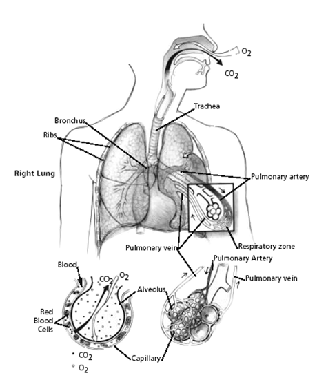
Red blood cells must be brought as closely as possible to the air we breathe, so that hemoglobin in the cells can give up waste carbon dioxide from cellular metabolism and take on oxygen. To accomplish this, the lungs have millions of tiny air sacs (alveoli) with very thin walls (alveolar membranes) containing microscopic blood vessels (capillaries) (see Figure 1 below). This anatomy of the lungs allows exposure of a large surface area of the blood to the air. Oxygen (O2) and carbon dioxide (CO2) gases diffuse (move) across the one cell thick alveolar membrane in opposite directions with the oxygen entering the blood and the carbon dioxide leaving it. This process is known as gas exchange.

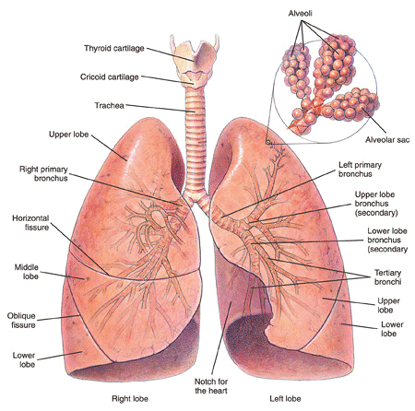
Figure 1: Bronchi and lungs.
The gas exchange part of the lungs is known as the lung parenchyma (see Figure 2 below). Air is delivered to the parenchyma of the lungs through the bronchial tree — a repetitively branching tubular system for air conduction. It consists of the trachea, from which arises a right and left main (primary) bronchus to the right and left lungs. Smaller bronchi branch from the main bronchus of the right or left lung, then to smaller bronchi to the various lobes of the lungs, then to even smaller bronchi (bronchioles) that eventually reach the alveoli.

Figure 2: Mechanism of gas exchange.
How Respiration Is Impaired
Many diseases can affect breathing. The most useful classification of respiratory disorders is based on the manner in which the ability of air to come into contact with the hemoglobin in red blood cells (RBCs) is disrupted.
There are only two ways in which respiration can be impaired, regardless of the exact nature of the disorder:
- Disease that prevents adequate amounts of air from reaching the gas-exchange level of the lungs (obstructive lung disease).
- Disease of the lung tissue itself that reduces gas exchange (restrictive lung disease).
Thus, physicians have found it broadly useful to classify respiratory disorders as obstructive or restrictive, or a combination or the two.
Chronic Obstructive Lung Disease (COPD)
Chronic obstructive pulmonary disease (COPD) is the most common type of lung disease seen by the Social Security Administration. “Obstruction” refers to the fact that air flow in and out of the lungs is impeded. The three most frequent types of COPD in adults are:
- Emphysema.
- Chronic bronchitis.
- Asthma.
In emphysema, lung tissue itself is destroyed. Damaged lung tissue forms non-functional spaces that trap air, and the lungs expand. The effect of these abnormalities is to obstruct air flow to and from the lungs. Emphysema and chronic bronchitis often occur together usually in people with a history of cigarette smoking.
Chronic bronchitis can also occur from long exposure to chemical fumes associated with a particular occupation. Inflammation of the inner surface of bronchi results from exposure to irritating substances. This inflammation, along with excessive secretion of bronchial mucous glands results in bronchial narrowing. Thus, bronchial tree resistance to air flow increases—there is obstruction of air flow.
Other less common causes of COPD include cystic fibrosis and bronchopulmonary dysplasia (BPD). These disorders, as well as asthma, have separate specific listings. See Can I Get Social Security Disability Benefits for Asthma? and Can I Get Social Security Benefits for Cystic Fibrosis?
Restrictive Lung Disease
The hallmark of restrictive lung disease is loss of usable lung volume, either due to:
- Disease of the gas exchange part of the lungs (lung parenchyma), or
- Some disorder outside of the lungs (extrapulmonary) that prevents air from adequately ventilating normal lung parenchyma.
Examples of Parenchymal Restrictive Lung Diseases
- Infections (bacterial, fungal, viral, parasitic). Chronic infections that damage lung tissue, such as severe bronchiectasis or advanced pulmonary TB, could result in some degree of restrictive impairment.
- Radiation, such as used for treatment of cancer, can damage lungs. Radiation lung damage is less of a problem than in the past, because modern equipment can direct therapeutic radiation in beams precisely delivered to the tumor. To the extent that this is impossible because of the size or location of the tumor, it is possible to have fibrotic lung damage caused by radiation.
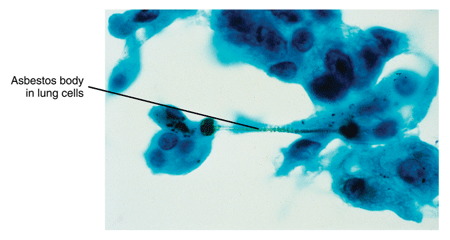
- Inhalation of damaging substances into the lung. Pneumoconiosis is a non-specific term that refers to lung damage from inhaling small particles of some kind. Examples of pneumoconiosis include that caused by asbestos (asbestosis) (see Figure 3 below), coal dust (anthracosis), beryllium (berylliosis) silicon dust (silicosis), aluminum, iron, tin (stannosis) and talc. Inhalation of toxic chemicals—such as acidic fumes—can also damage lung tissue.

Figure 3: Asbestos in lung cells.
- Drug side-effects.
- Autoimmune diseases. Autoimmune disorders such as sarcoidosis, scleroderma, systemic lupus erythematosus (SLE), rheumatoid arthritis (RA), polymyositis, ankylosing spondylitis, and mixed connective tissue disorders can cause pulmonary disease. In all of these disorders, some kind of immune system dysfunction damages the lungs. See Can I Get Social Security Disability Benefits for Lupus? and Can I Get Social Security Disability Benefits for Arthritis and Joint Damage?
- Idiopathic diffuse interstitial pulmonary fibrosis (cryptogenic fibrosing alveolitis). This condition causes progressive scarring of the lungs on a microscopic level. This damage can result in decreased gas exchange capacity of the lungs, increasingly severe hypoxemia (lack of oxygen in the blood) and eventually death from respiratory failure. The rate of progression is highly variable, but median survival is less than 3 years. Some cases remain slowly progressive for a number of years, then are triggered by some unknown event to become rapidly more severe.
- Other diseases. Examples of other diseases that can cause pulmonary fibrosis include alveolar proteinosis, acquired immunodeficiency syndrome (AIDS), acute respiratory distress syndrome (ARDS), amyloidosis, bone marrow transplantation, cancer, eosinophilic granuloma, eosinophilic pneumonia, lipoid pneumonia, genetic metabolic diseases caused by enzyme deficiencies (Gaucher’s disease, Niemann-Pick disease), Hermansky-Pudlak syndrome, pulmonary vasculitis, tuberous sclerosis, and neurofibromatosis. See Can I Get Social Security Disability Benefits for HIV / AIDS?
Examples of Extrapulmonary Restrictive Lung Diseases
- Abnormal spinal curvatures. Abnormal curvatures of the spine can interfere with normal breathing movements. Scoliosis is a common spinal disorder, but will not compromise breathing until the major abnormal curve reaches about 60 degrees. Kyphosis is an abnormal curvature of the upper (thoracic) spine that causes a bent-forward or “hunched-over” posture. Kyphoscoliosis is a combination of both kyphosis and scoliosis.
- Surgical resection. Removal of lung tissue limits the surface area available for gas exchange.
- Spondyloarthropathies. Inflammatory disorders of the spine, such as ankylosing spondylitis, can make the spine more rigid by increased calcification of the spine and associated soft tissue ligaments. The ribs attach to the spine and their movement is impeded during expansion and relaxation of the chest during breathing. This limitation will decrease breathing capacity. See also Can I Get Social Security Disability Benefits for Back Pain?
- Thoracoplasty. Thoracoplasty, usually involving removal of one or more ribs, distorts the normal shape of the chest. The intercostal muscles between the ribs help expand the chest during inspiration. However, mechanical distortion of the chest wall is probably more important in decreasing ability to ventilate the lung during respiratory movements of the chest.
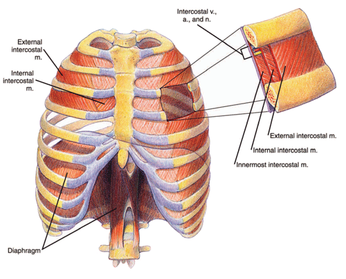
- Obesity. Marked obesity can result in a significant reduction in ventilation capacity. Because of the weight of fat on the chest wall, the work of breathing is increased during inspiration. Furthermore, abdominal subcutaneous fat as well as intra-abdominal fat resists movement of the diaphragm—a respiratory muscle consisting of two sheets of muscle separating the chest and abdominal cavities (see Figure 4 below). The diaphragm must be able to move downward with inhalation in order to maximally expand the chest.

Figure 4: Chest cavity and diaphragm.
- Other important causes of restrictive respiratory impairment. Many disorders can result in weakness affecting the muscles of respiration (diaphragmatic muscles or intercostal muscles). Myasthenia gravis is an autoimmune disease that causes weakness through a biochemical interruption of the body’s ability to excite muscles, including the muscles of respiration. In fact, respiratory failure is usually the cause of death in myasthenia. There are a large number of muscle diseases (myopathies) that can affect respiration, although fairly rare in the general population.
Strokes and Breathing Problems
Strokes (cerebrovascular accidents, CVAs) are frequently adjudicated by the Social Security Administration. See Can I Get Social Security Disability Benefits After a Stroke? However, most of the time the treating source—even if a neurologist—does not consider the possibility of a breathing deficit resulting from the stroke. However, paralysis of half of the diaphragm is a definite possibility with resultant decreased ability to ventilate the lungs. Because treating sources, or consulting neurologists paid by the Social Security Administration, usually do not specifically address the possibility of diaphragmatic paralysis, SSA adjudicators are also likely to not even think of the possibility. Claimants themselves, following a stroke, may have thinking difficulties and neurological problems that occupy their attention as well as the attention of their family. The claimant cannot be counted on to mention breathing difficulty—especially since he or she may not even have symptoms in a resting state. The Social Security Administration adjudicator should ask to treating doctors, especially neurologists, regarding possible breathing deficits in post-stroke patients.
Pulmonary Function Studies
Pulmonary function study (PFS) is a general term that applies to any type of respiratory testing. The basic types of PFS are:
Shortness of breath is one of the most common allegations by claimants seeking Social Security disability benefits. The Social Security Administration pays for many PFS tests (especially spirometry) to address allegations of shortness of breath.
The results of these tests determine whether you qualify for disability benefits under a listing. See Winning Social Security Disability Benefits for Lung Disease by Meeting a Listing.
No pulmonary function study is more useful or more frequently performed than spirometry. Spirometry is the most important test for evaluating the severity of obstructive pulmonary disease. Spirometry requires you to inhale then exhale into a device called a spirometer. The device measures the volume of air that you can inhale and exhale and displays the result as a breathing curve on a graph called a spirogram.
If you have lung disease, but have not had a spirometry test, the Social Security Administration may arrange for you to have one. Even if you have had the test, the Social Security Administration may require you to be retested because the test must be administered in accordance with strict rules. Accurate testing must be done to assure you are treated fairly.
The test results must include the actual breathing curves. Spirometry test results in medical records often do not include the actual breathing curves, just the numerical results.
If there is no clinical evidence that you have significant lung disease and the reported spirometric values in your medical records are not significantly abnormal, the Social Security Administration will not send you for retesting. In fact, the Social Security Administration is not obligated to obtain spirometry on claimants who have no evidence in their medical records of a respiratory disorder, even if there are no spirometric values of any kind in the file.
Arterial Blood Gas Study (ABGS)
ABGS is the most important test used for the evaluation of the restrictive pulmonary disorders that involve a decrease in gas exchange—the parenchymal restrictive lung diseases. ABGS is performed on a sample of blood from the radial artery in the wrist or brachial artery in the arm, unlike most blood samples that are taken from a superficial vein just under the skin.
An ABG test checks how well your lungs are doing in moving oxygen into the blood and removing carbon dioxide from it. ABGS measures arterial oxygen pressure (PaO2), also known as oxygen tension, carbon dioxide pressure (PaCO2), and acidity (pH).
The Social Security Administration should not send you for ABGS, unless absolutely necessary to determine whether you are disabled, because it is invasive. Although complications are unusual, the person sticking a needle in the radial artery could damage the artery or other structures in the wrist; use of the brachial artery carries additional risk and should be done only when the radial artery cannot be used. The needle stick is also painful in either method.
Carbon Monoxide Diffusing Capacity (DLCO)
DLCO is a test used to evaluate of the severity of parenchymal restrictive lung diseases. It is of little value in assessing severity in COPD. The test measures how well your lungs can transfer carbon monoxide (CO) into the blood. By measuring how easily CO moves across the alveolar membrane of the lungs, doctors can deduce whether there is also a problem limiting the exchange of oxygen and carbon dioxide between blood and the atmosphere.
Two different methods are used for this test. The single-breath method is the one required by the Social Security Administration. This method requires you to take a breath of air containing a very small amount of carbon monoxide from a container while measurements are taken. (The other method, the steady-state method, requires you to breathe air containing a very small amount of carbon monoxide. The amount of carbon monoxide in the breath you exhale is then measured.)
DLCO can be abnormal in emphysema, if the disease is very advanced, because lung tissue is damaged. However, DLCO is not an accurate enough means of determining the severity of emphysema; spirometry is more appropriate. In some instances, more applicable to clinical medicine than disability determination, DLCO testing can help distinguish between emphysema and asthma, because asthma will have normal values.
Continue to Winning Social Security Disability Benefits for Lung Disease (Chronic Pulmonary Insufficiency) by Meeting a Listing.
